If you point to pain just above your buttock, you might immediately think, “My lower back hurts.”
That's understandable.
The problem is that the area around the low back, pelvis and hip contains several structures that can produce overlapping symptoms. One of those structures is the sacroiliac, or SI, joint.
That doesn't mean every ache near the buttock is SI joint pain. In fact, identifying the SI joint as a pain source can be challenging. A complete history and physical examination are important for differentiating it from other causes of low back and buttock pain.
Where is the SI joint?
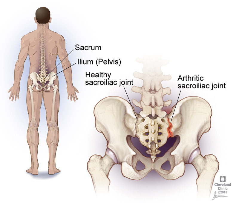
The sacroiliac joints connect the pelvis to the sacrum, which sits at the base of your spine.
They help transfer loads between your trunk and legs.
SI joint-related pain is often felt around the buttock or just to the side of the lower back. It can sometimes extend into the thigh, hip or groin.
But location alone isn't enough to confirm that the SI joint is responsible.
Why can it feel like ordinary back pain?
The low back, pelvis and hip work together during activities such as walking, climbing stairs, getting out of a chair, running and lifting.
Because these areas share movement and load, symptoms can overlap.
For example, pain near the back of the pelvis could potentially involve the SI joint, lumbar spine, hip or surrounding muscles and connective tissues.
That's why simply pressing on one painful spot isn't a complete diagnostic strategy.
How do clinicians evaluate the SI joint?
A clinician may look at your walking pattern, strength, hip mobility, lumbar movement and how different movements reproduce your symptoms.
There are also specific SI joint provocation tests.
Interestingly, individual tests aren't particularly reliable when used by themselves. A combination of several provocation tests provides more useful diagnostic information. A commonly referenced clinical approach uses a cluster of five tests, with three or more reproducing the patient's familiar pain supporting an SI joint pain diagnosis.
That illustrates an important principle:
Good clinical reasoning usually comes from a pattern of findings, not one test.
What about an MRI?
An MRI can show anatomy, but imaging doesn't always identify the exact source of SI joint pain.
Imaging may be useful when a clinician is considering other conditions or when the result could change management. It isn't automatically necessary simply because someone has pain near the SI joint.
What can treatment look like?
When SI joint-related pain is suspected, conservative care may include education, exercise and manual therapy depending on the individual's presentation.
The goal isn't simply to “put the SI joint back in place.”
Instead, treatment should address the person's symptoms, movement, strength, activity demands and ability to tolerate load.
For some people, improving pelvic or hip strength may be relevant. For others, changing how they move, temporarily modifying aggravating activities or addressing mobility may be more appropriate.
There isn't one SI joint exercise that everyone needs.
The important takeaway
Pain near your SI joint doesn't automatically mean your SI joint is the problem.
And pain in the low back doesn't automatically mean the lumbar spine is the only area worth examining.
The pelvis, hips, lumbar spine and surrounding tissues all contribute to movement.
A good evaluation looks at the entire picture before deciding what deserves attention.